Benign migratory glossitis

This visual ‘atlas’ will help you to identify and describe non-malignant, potentially malignant and malignant oral lesions.

A diverse range of mucosal lesions can occur in the oral cavity. The history and clinical presentation of the non-malignant conditions listed below will help you differentiate them from potentially malignant or cancerous lesions. However, occasionally this can be difficult. If a lesion is suspicious, refer your patient for biopsy and definitive diagnosis.

The stage when oral cancer is diagnosed is important, as it indicates prognosis. Stage I and II oral cancer corresponds directly to size, with no evidence of metastases. Stage III and IV oral cancer has spread to the lymph nodes or distant organs. Detection of oral cancer at an early stage can save your patient’s life.

Erythema migrans

Geographic tongue

Chronic inflammatory condition of the tongue. Filiform papillae (responsible for giving the tongue its texture and the sensation of touch) on the dorsal surface of the tongue are lost then return, in a recurring pattern across different areas. These patterned areas on the tongue are slightly depressed and red, with a surrounding white rim – giving the appearance of a topographical map, with changes week by week.

Occurs in around 2% of the population and is mostly asymptomatic, although some patients note increased sensitivity to acidic and spicy foods. These periods of sensitivity may occur when patients are stressed or ill. Asymptomatic patients require no treatment.
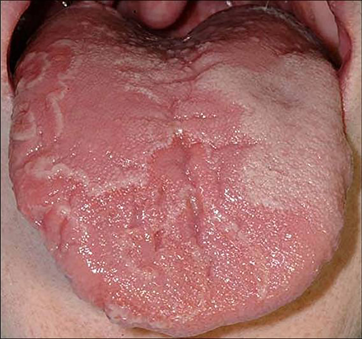
Areas of slight depapillation of the dorsal surface of the tongue, which are slightly depressed and erythematous surrounded by grey-white areas.

Last updated: March 2021