Generalised erythema (redness), desquamation (skin peeling) and oedema of the gingiva usually effecting the whole of the attached gingivae. Desquamative gingivitis is a clinical term describing the appearance. The cause of desquamative gingivitis is most often oral lichen planus, but can also be the clinical appearance of a vesiculobullous condition (fluid filled lesions) such as pemphigoid or pemphigus.
Patients will often experience pain with tooth brushing, leading to plaque-induced gingivitis and periodontitis. However, this condition needs to be recognised separately and the underlying cause diagnosed and treated. Enhanced oral hygiene alone will not resolve desquamative gingivitis and will lead to continued persistence, pain and suffering.

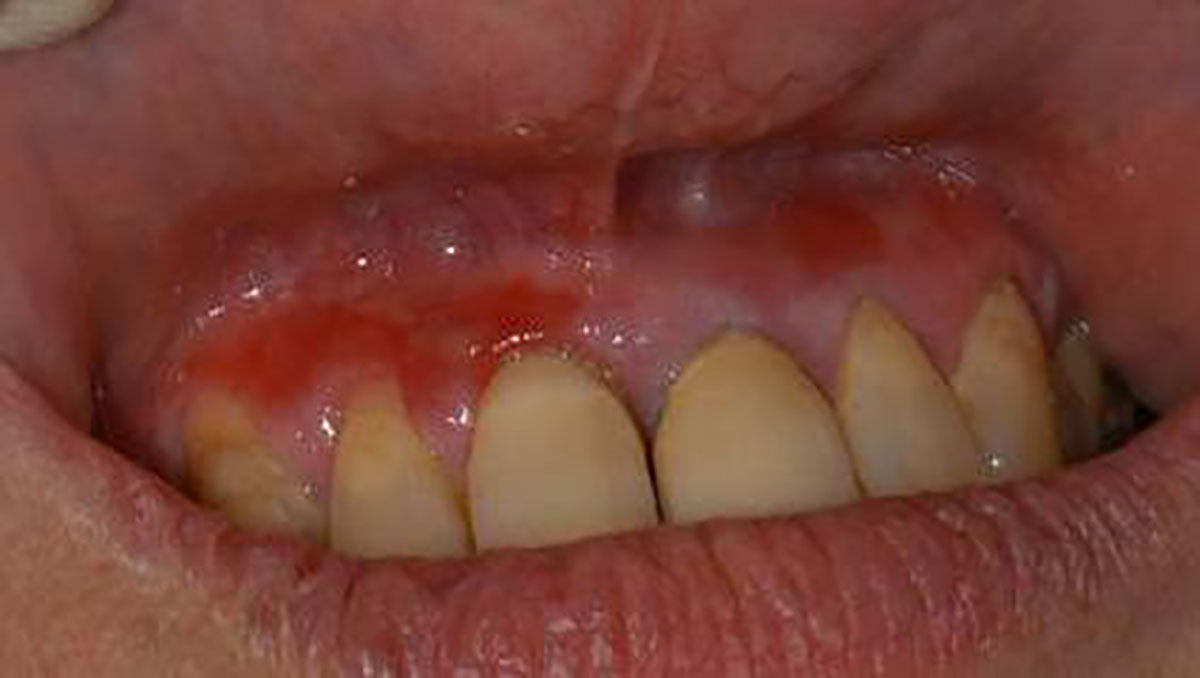
Painful area of erythema of the gingivae extending from the mesial of the upper right central to the gingival margin of the upper right canine. A small erythematous area is also present apical to the upper left lateral incisal gingival margin. These areas were very painful to touch, making toothbrushing very difficult.

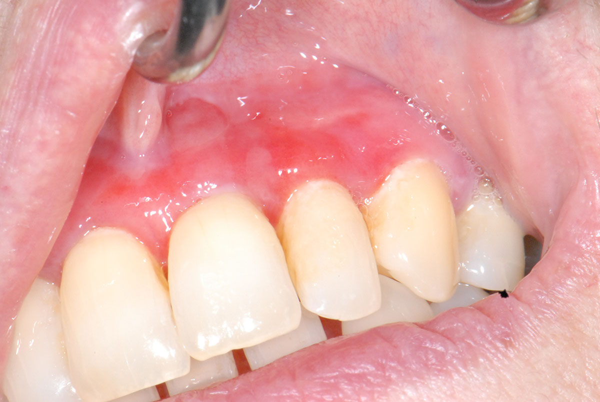
Erythema and ulceration across the labial maxillary gingivae extending into the labial sulcus. Very little plaque present.

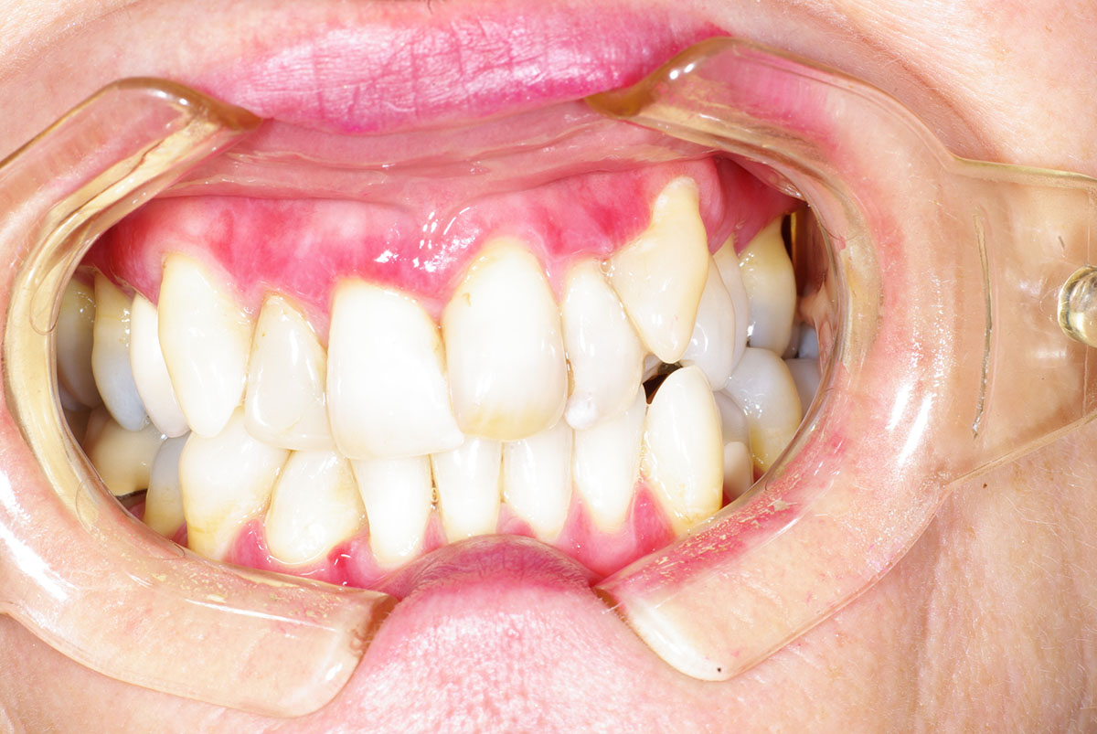
Erythema with faint striae on almost all the labial gingivae but separate from the free gingival margin that appears normal.

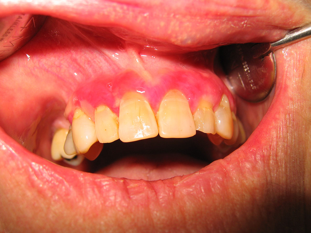
Extensive erythema across the anterior maxillary labial gingivae with areas of ulceration particularly around the upper right canine