Primary herpetic gingivostomatitis
Recurrent herpes labialis (cold sores)
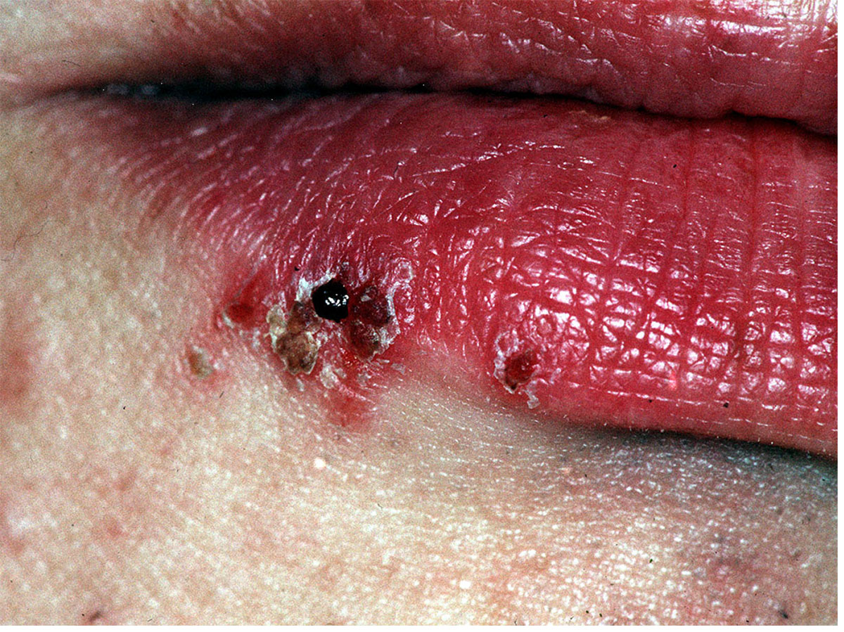
Secondary infection with herpes simplex virus type 1 (HSV-1) that effects the vermillion of the lip with recurring sores following a pattern of slight paraesthesia, redness, appearance of vesicles, superficial ulceration, crusting and eventual healing over a period of 5 to 7 days. HSV-1 resides in the trigeminal ganglion latently after the initial primary infection, primary herpetic gingivostomatitis, that causes a widespread infection with fever, malaise, lethargy and widespread ulceration of the oral mucosa and gingivae over a period of 7 to 10 days. This primary infection mostly occurs in infants, when it is not as severe, but can be seen following oral sex in a seronegative young adult. Recurrent herpes labialis occurs in about 25-30% of individuals and is usually precipitated by concomitant illness, exposure to sunlight or wind and in immunocompromised individuals when it can become severe and prolonged.

Crusting ulcers on the right lower lip at the junction of the vermilion and skin that were previous vesicles that burst several days before this presentation.