Human papillomavirus (HPV)

This visual ‘atlas’ will help you to identify and describe non-malignant, potentially malignant and malignant oral lesions.

A diverse range of mucosal lesions can occur in the oral cavity. The history and clinical presentation of the non-malignant conditions listed below will help you differentiate them from potentially malignant or cancerous lesions. However, occasionally this can be difficult. If a lesion is suspicious, refer your patient for biopsy and definitive diagnosis.

The stage when oral cancer is diagnosed is important, as it indicates prognosis. Stage I and II oral cancer corresponds directly to size, with no evidence of metastases. Stage III and IV oral cancer has spread to the lymph nodes or distant organs. Detection of oral cancer at an early stage can save your patient’s life.

Verruca vulgaris

Squamous papilloma

Benign, small isolated outgrowths of the oral mucosa that can have small finger-like projections resulting in a rough cauliflower-like surface. These are caused by non-oncogenic sub-types of human papilloma virus (HPV), usually 1, 6, 11 or 57. Acquired by direct contact or self-inoculation. These growths can be removed if unsightly or interfering with good oral hygiene.
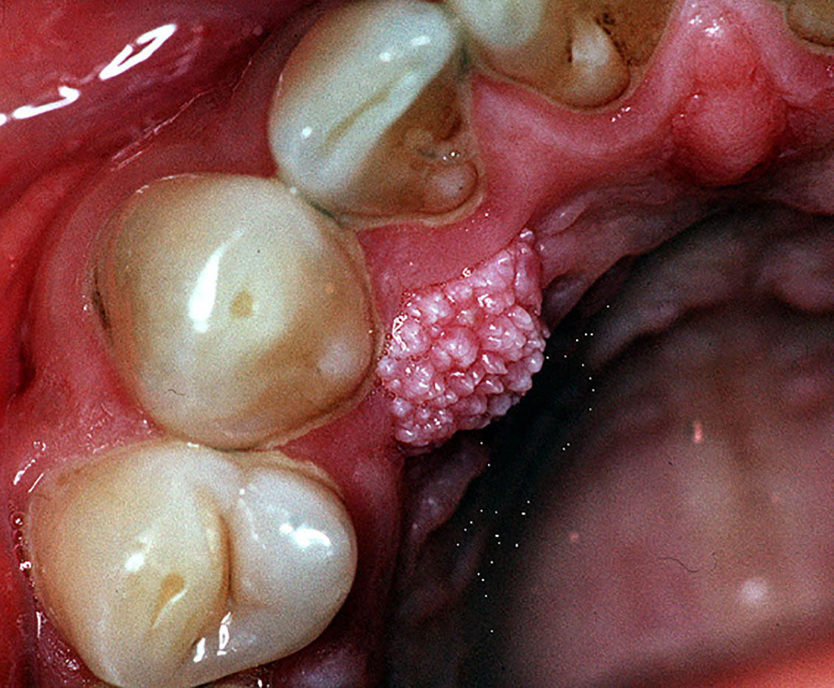
An exophytic mass on the palatal mucosa close to the gingival margin of the upper right canine that has a rough cauliflower-like surface. This was asymptomatic and been present for many years.

Last updated: March 2021