Pemphigus vulgaris
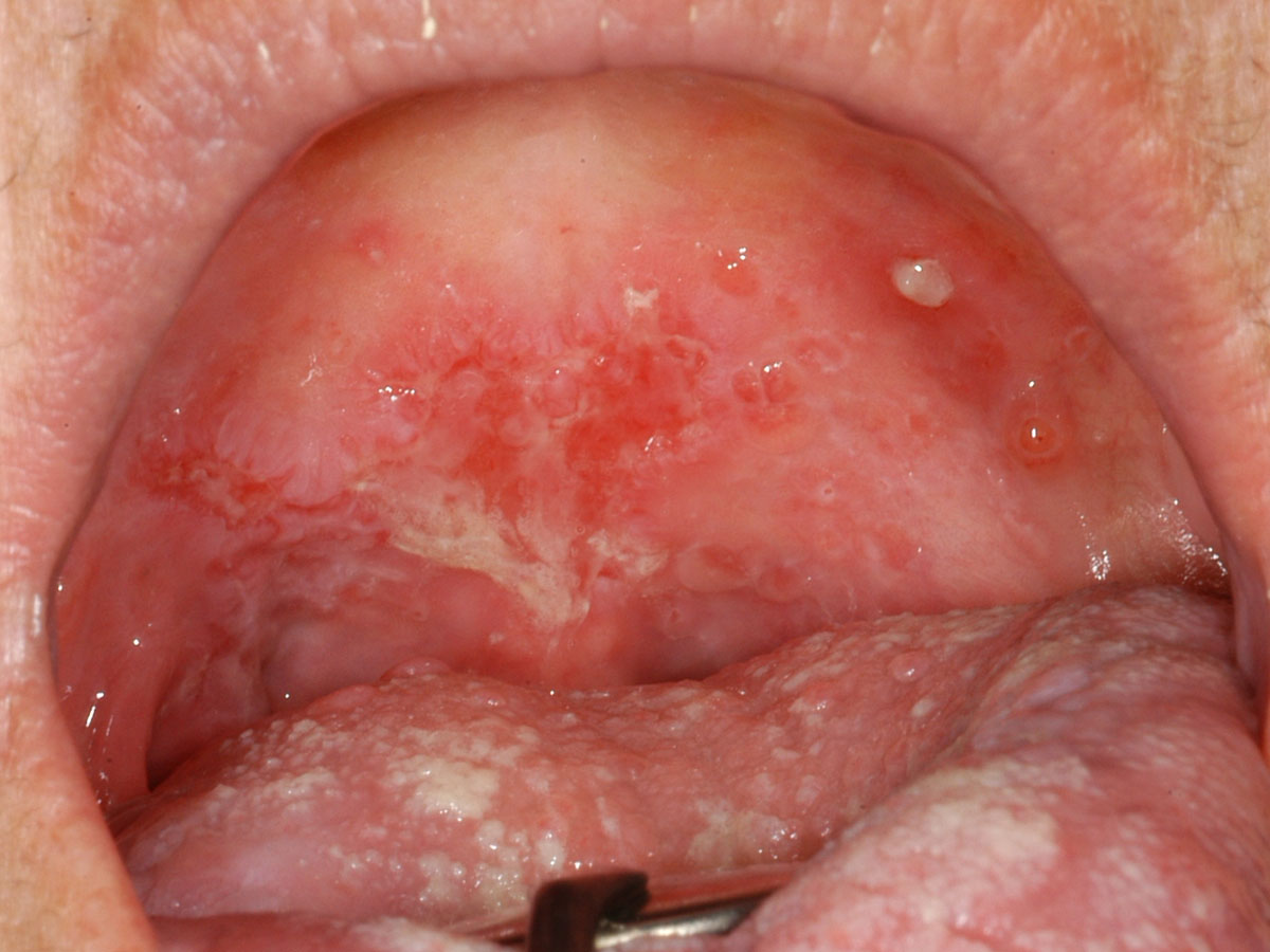
Pemphigus vulgaris is the most common autoimmune intra-epithelial bullous disease that is also potentially life-threatening. It causes blisters on the skin and/or mucosa. Although this is a rare condition, the mouth is affected in about 90% of people and in some patients it will be the only site affected. Accompanying oral lesions are poorly defined, irregular persistent ulcers.

Extensive painful erythema, ulceration and deflated bullae across the soft palate in this patient with pemphigus vulgaris.
Mucous membrane pemphigoid
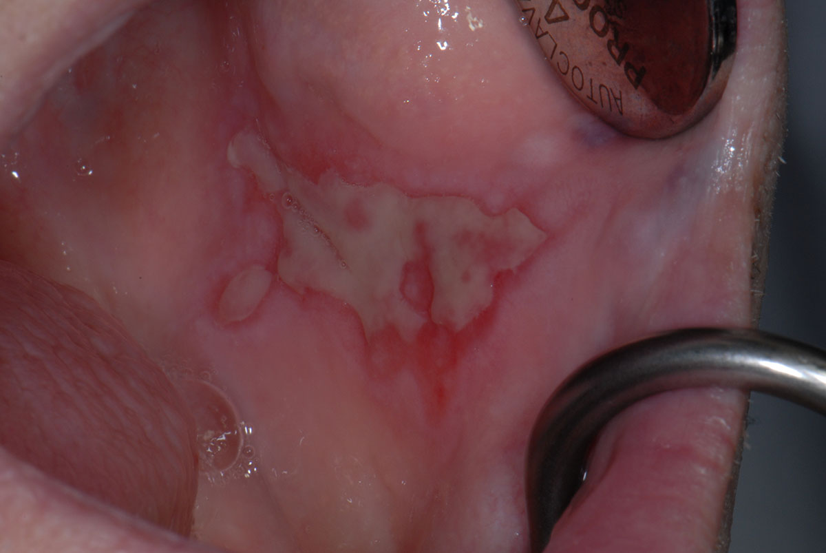
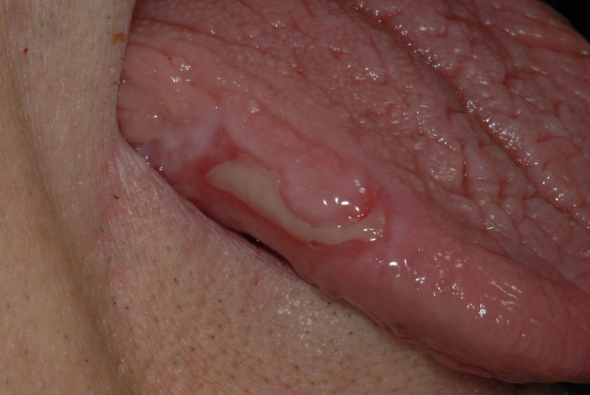
A rare mucosal autoimmune sub-epithelial blistering disease, with a wide variation in severity: from mild, almost painless oral ulceration, to severe blisters with scarring affecting the mouth, larynx, oesophagus and conjunctiva which can lead to blindness.

Painful ulceration across the left buccal mucosa surrounded by bright erythema

Deflated bullae that has ulcerated with surrounding erythema on the right lateral margin of the tongue in the same patient as imaged above.