Homogenous leukoplakia
Non-homogenous leukoplakia
Verrucous leukoplakia
Nodular leukoplakia
Erythroleukoplakia
Leukoplakia are white plaques that are not caused by any other condition and which have an increased potential for malignant transformation. This is the most common potentially malignant oral mucosal condition.
Potential for malignancy has been reported to be around 1-7% for homogenous leukoplakia, and 5-50% for non-homogenous leukoplakia. While the cause is unknown, risk factors include smoking, drinking alcohol and betel quid chewing.
Diagnosis includes a biopsy for assessment of the presence of dysplasia. Management involves reducing behavioural risks, e.g. cessation of smoking and alcohol. Depending on clinical and histopathological appearance, treatment may involve excision and will require long-term monitoring of all oral mucosa.
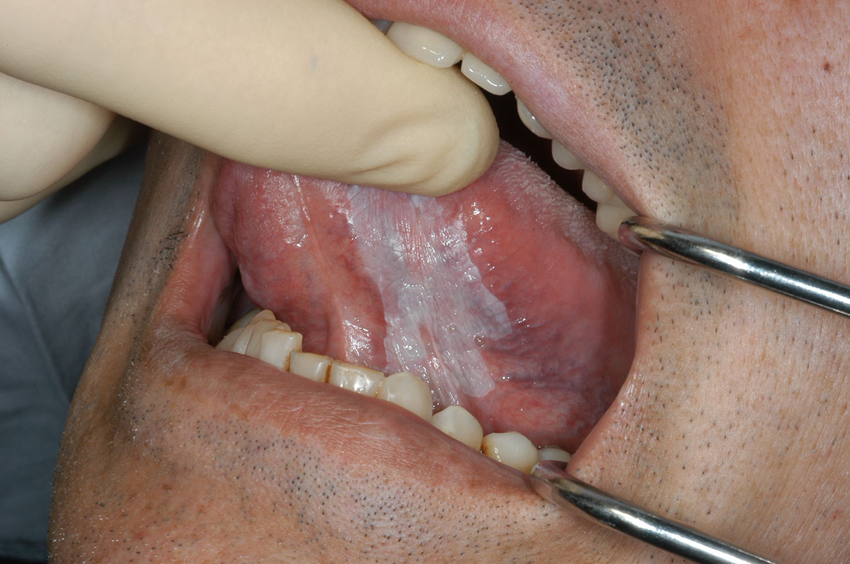
A flat homogeneous white plaque present on the ventral surface of the left tongue, extending onto the floor of mouth. This lesion was asymptomatic and only observed on routine examination in a heavy smoker.