Reticular oral lichen planus
Erythematous oral lichen planus
Ulcerative oral lichen planus
Plaque-like oral lichen planus
A chronic, immune-mediated mucosal condition occurring in less than 1% of the population, and more often in middle age and older. Oral lichen planus has a variable clinical appearance with a reticular pattern, the most common consisting of striae bilateral buccal mucosa that can extend to the lateral margins of the tongue. It can also appear as areas of erythema, ulceration and plaque-like regions, or a combination of all of these.
Similar appearance can be observed with drug-associated hypersensitivity reactions, contact hypersensitivity to restorative materials and graft versus host disease.
Symptomatic treatment requires topical anti-inflammatory agents, while asymptomatic lesions require no treatment. Long-term monitoring is advisable as there is a potential for around 1% to undergo malignant transformation.
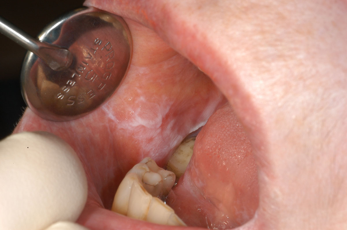
White striae extending across the right buccal mucosa in a reticular pattern with very little erythema and no ulceration.
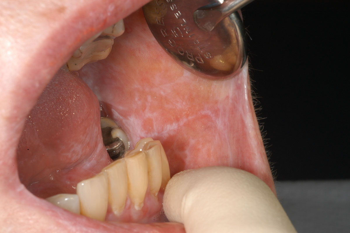
White striae extending across the left buccal mucosa in a reticular pattern with very little erythema and no ulceration. This is the same patient as in image above, who presented with bilateral buccal mucosal asymptomatic oral lichen planus.
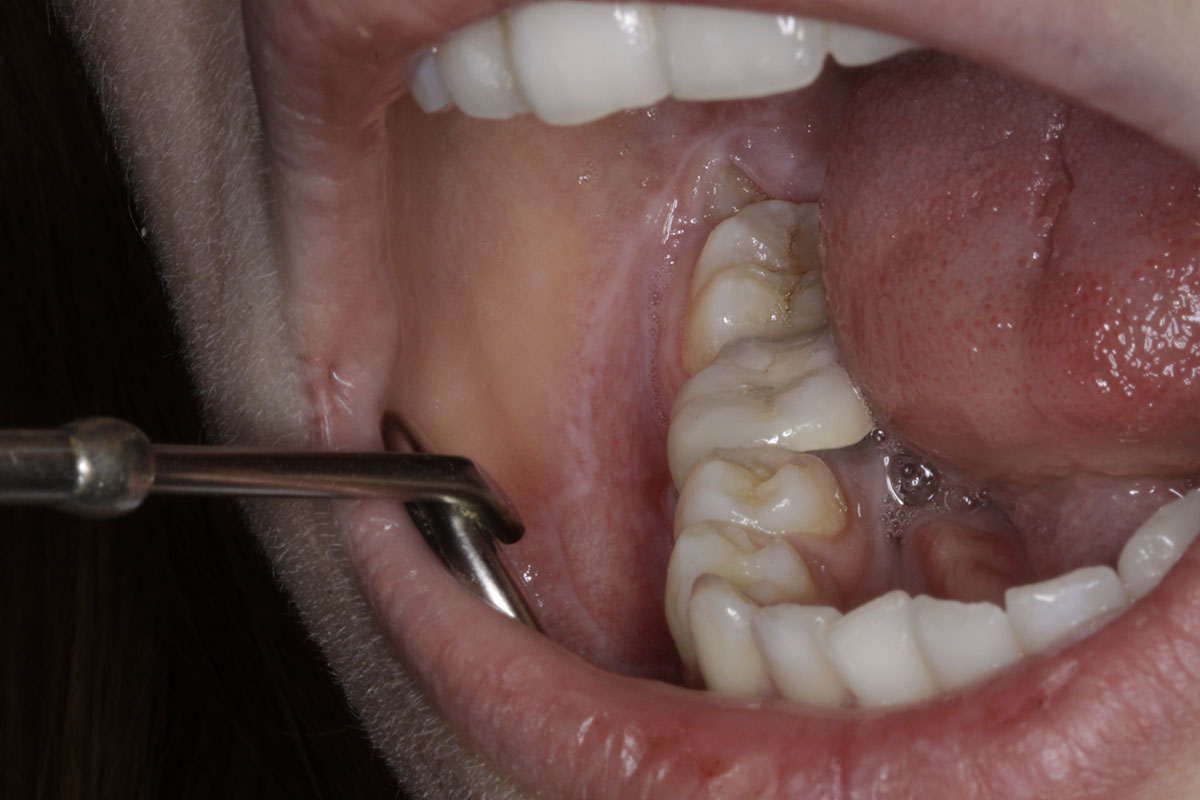
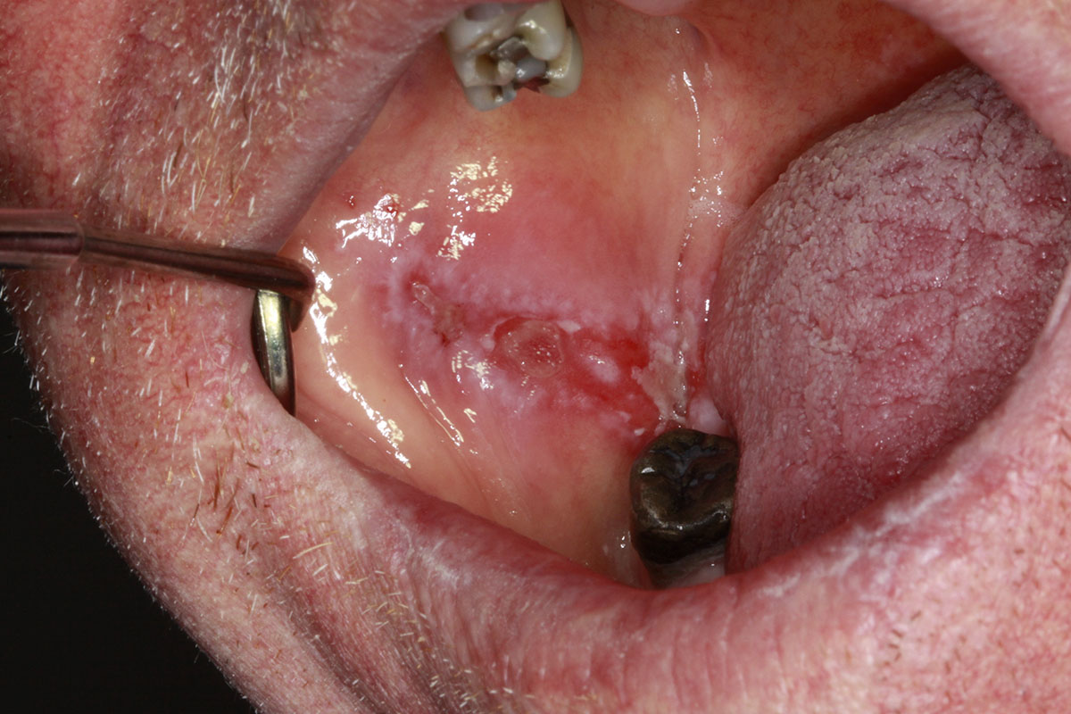
Striae extending to the depth of the mandibular sulcus on the right buccal mucosa and posteriorly onto the tissue overlying the erupting third molar. This patient’s symptoms were associated with the erupting third molar, not lichen planus.
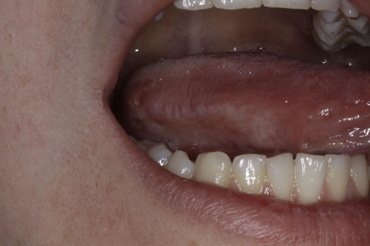
Striae on the right lateral margin of the tongue with no erythema or ulceration in the same patient as in image above.
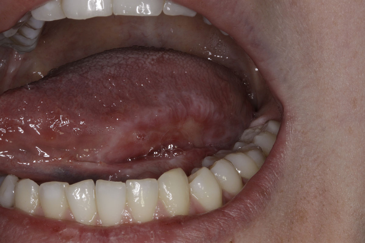
An area of striae with erythema and ulceration on the left lateral margin of the tongue in the same patient as in image above. This ulcerated area had been present for several months and was painful to touch. Biopsy of the non-ulcerated region on this site was diagnostic of oral lichen planus.
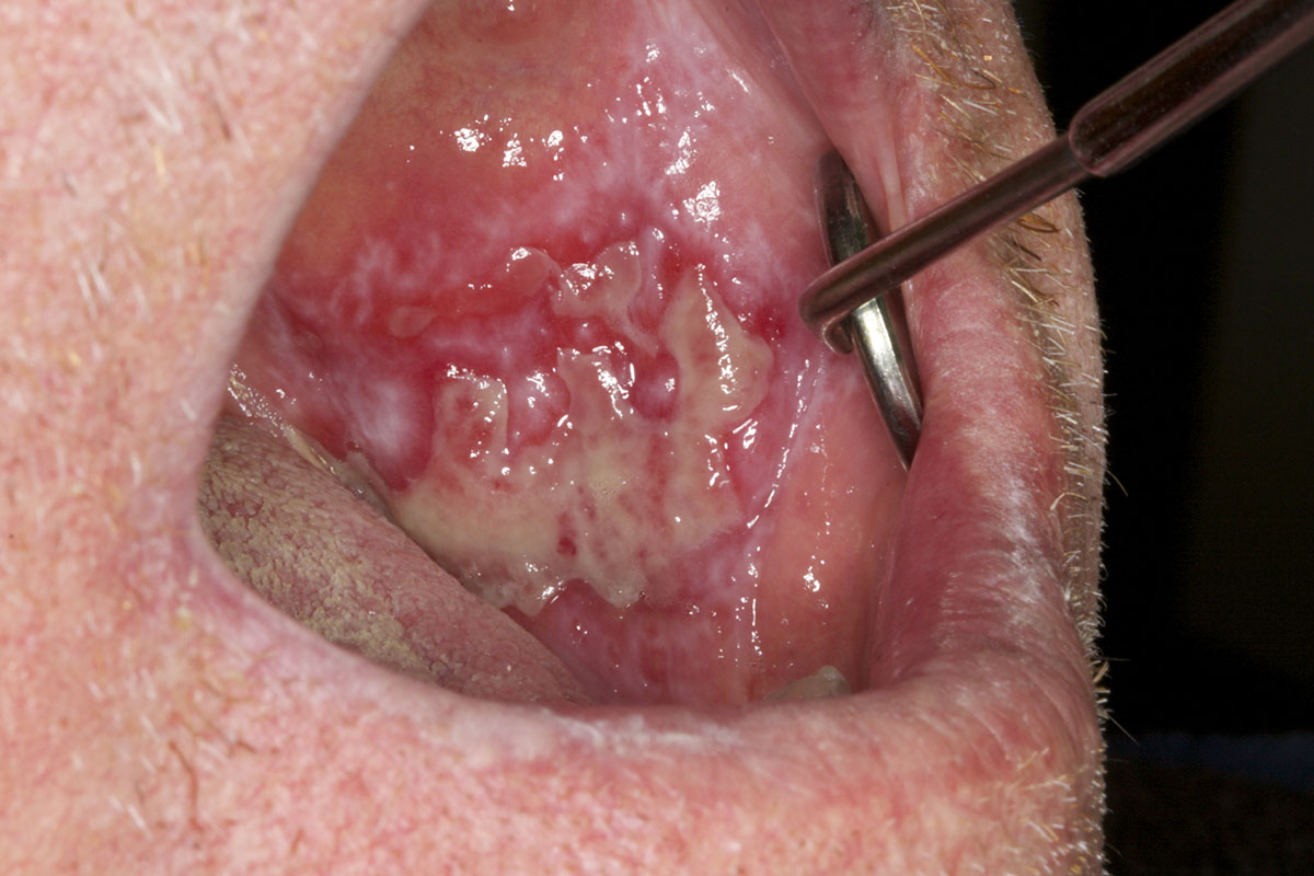
Extensive ulceration, erythema and striae on the right buccal mucosa that was very painful.
Painful ulceration with erythema and surrounding striae on the right buccal mucosa. This is the same patient as in image above. Extensive mucosal changes were diagnosed as histopathological as oral lichen planus.